
Bicuspid aortic valve in sport: assess the valve and the aorta
Bicuspid aortic valve is not one risk category. Sport decisions need valve function, aortic dimensions, blood pressure, symptoms and shared review.
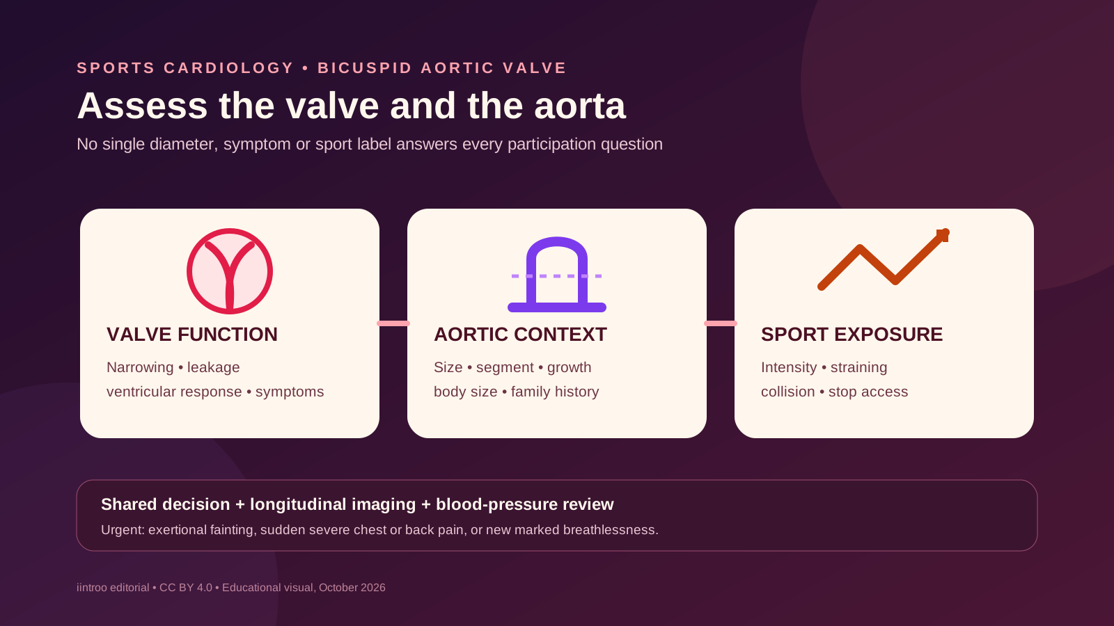
Key points
- A bicuspid aortic valve can be associated with narrowing, leakage and enlargement of the ascending aorta; each feature needs separate assessment.
- Current AHA/ACC guidance supports individualised participation after complete evaluation rather than treating every athlete with BAV as the same.
- Exertional chest pain, fainting, sudden severe chest or back pain, new marked breathlessness or a rapid decline needs urgent assessment.
Separate the valve diagnosis from its consequences
This guide is for athletes with a bicuspid aortic valve (BAV), their families, coaches and sport medical teams. BAV means the aortic valve formed with two functional leaflets rather than the usual three. Some people have normal flow for years; others develop aortic stenosis, regurgitation or enlargement of the aortic root or ascending aorta. The diagnosis therefore does not produce one training limit. Decisions start with symptoms, examination, blood pressure, ECG and imaging that describes both valve function and the aorta.
The 2025 AHA/ACC scientific statement organises BAV with aortopathy separately from other inherited aortic conditions and emphasises shared decision-making. It notes that aortic dissection in athletes with BAV-associated aortopathy is uncommon, so participation may be reasonable after complete size assessment and risk stratification. 'Uncommon' is not 'impossible', and a normal-feeling athlete can still have important valve obstruction or enlargement. The athlete needs scheduled cardiology surveillance even when training is going well.
Measure size, change and context rather than chasing one number
The cardiology team considers the aortic diameter, the body-size-adjusted value where appropriate, which segment is enlarged, image quality and whether the measurement is changing. Family history of aortic dissection, coarctation, hypertension, genetic features and pregnancy plans can alter the conversation. Echocardiography is central, while CT or MRI may be needed when the full aorta is not well seen or measurements need confirmation. Comparing studies requires consistent technique; a small apparent change can reflect measurement variation rather than true growth.
Guideline tables provide ranges that help structure discussion, but they are not do-it-yourself clearance charts. Sex labels, body size, age, growth and the limitations of sparse athlete outcome data all matter. Aortic dimensions in trained athletes can overlap with disease-associated enlargement, yet BAV still requires disease-specific review. The 2022 ACC/AHA aortic-disease guideline recommends lifelong surveillance when indicated and aggressive attention to blood pressure. An isolated online number cannot replace longitudinal imaging interpreted by the treating specialist.
Map cardiovascular findings to real sport demands
A sport review should describe dynamic intensity, heavy static effort, collision, acceleration, breath-holding, competition stress and the ability to stop safely. Very heavy resistance work can create large transient blood-pressure loads, while endurance sport creates sustained volume load. The relevance depends on valve severity, aortic size and the athlete's response, not on a simple endurance-versus-strength label. Clinician-supervised exercise testing can help assess symptoms, blood pressure, rhythm and capacity at a workload that resembles training.
Training plans may use moderated intensity, avoidance of maximal straining or another tailored limit, but these are clinical decisions rather than universal prescriptions. Coaches should receive functional boundaries and warning signs with the athlete's consent, not a copy of the entire medical record. If a restriction changes, document who made the decision and when review is due. Performance pressure, scholarships or selection deadlines should not replace informed consent, and a normal smartwatch trace does not evaluate the valve or ascending aorta.
Keep surveillance and an emergency route visible
Stop exercise and seek urgent assessment for exertional fainting, chest pressure, a sudden drop in capacity, new disproportionate breathlessness or sustained palpitations. Sudden severe chest, back or abdominal pain, especially with collapse, weakness or neurological symptoms, requires emergency services. These symptoms have many possible causes and should not be self-diagnosed as the valve or aorta. Venues should still have a rehearsed emergency action plan and access to an automated external defibrillator; surveillance cannot eliminate every event.
Follow-up intervals depend on valve function, aortic size and change, age, treatment and clinical context. Pregnancy, new hypertension, a major symptom, a family diagnosis or a change to higher-intensity sport should trigger review rather than waiting for the next routine appointment. This article does not interpret an echo, set a lifting limit, clear competition or recommend surgery or medicine. Those decisions require a cardiologist familiar with aortopathy and sport, using current images and the athlete's own goals.