
Inflammatory myopathy in athletes: exercise supports care but does not replace disease control
Dermatomyositis and other inflammatory myopathies can affect muscle, swallowing, lungs and heart. Tailored exercise belongs inside specialist disease management.
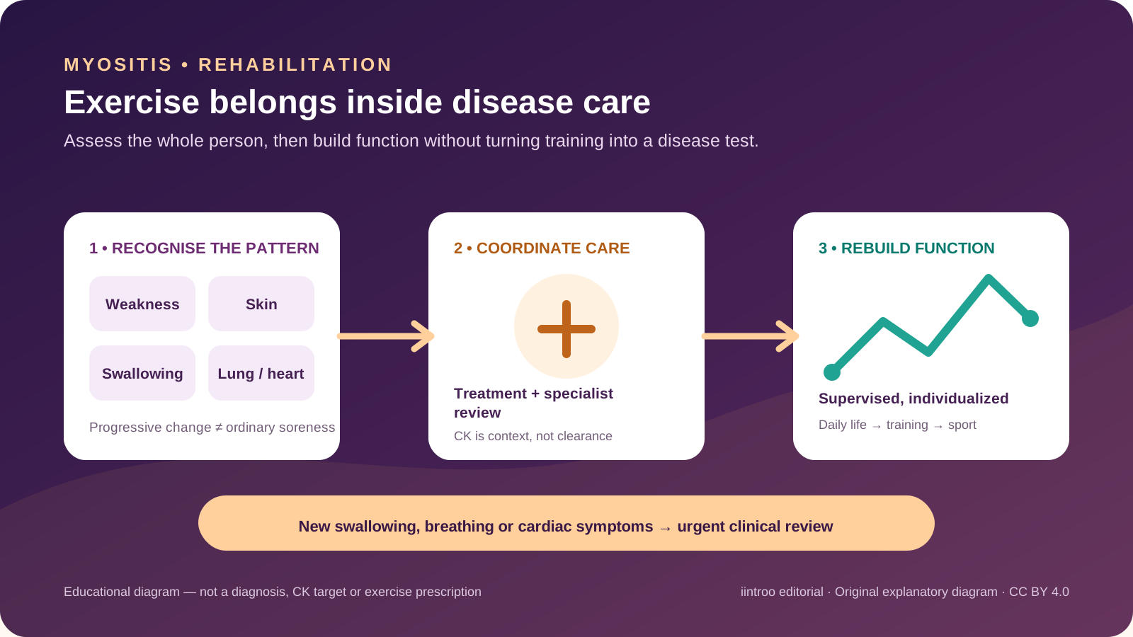
Key points
- Progressive proximal weakness, swallowing change, rash or breathing symptoms need medical assessment; a raised or normal CK alone does not define the condition.
- Guidelines encourage individualized exercise and rehabilitation across disease activity, coordinated with specialist treatment and safety assessment.
- Trials suggest gains in strength and aerobic capacity, but samples are small, diagnoses are heterogeneous and programmes are not universal prescriptions.
Recognise weakness that is not ordinary post-training soreness
This guide is for people with suspected or diagnosed idiopathic inflammatory myopathy, including dermatomyositis, immune-mediated necrotizing myopathy, overlap myositis and selected cases historically labelled polymyositis. These are rare, heterogeneous autoimmune conditions rather than one training injury. Difficulty rising from a chair, climbing stairs, lifting the arms, holding the head up or repeating previously easy tasks may be more important than pain. Dermatomyositis can add characteristic skin changes. Fatigue or delayed soreness after a hard session alone does not establish inflammation.
Seek prompt assessment for progressive weakness, a new rash with weakness, dark urine, persistent fever or a major loss of function. Trouble swallowing, choking, a weak cough or new shortness of breath needs urgent clinical advice because the swallowing muscles, lungs or respiratory muscles may be involved. Chest pain, fainting, marked palpitations or severe breathlessness requires urgent care. Coaches should stop loading the affected movement and support medical evaluation rather than prescribing recovery supplements, massage or another maximal test.
Assess a multisystem disease, not one enzyme value
Diagnosis combines the pattern of weakness, examination, muscle enzymes, autoantibodies and selected imaging, electrical testing or biopsy. Creatine kinase can be markedly raised, only mildly changed or occasionally normal depending on subtype, activity and timing. Recent strenuous exercise can also raise CK. The result therefore cannot be used by an athlete as a stand-alone disease or clearance score. The specialist may assess skin, joints, swallowing, nutrition, lung function and imaging, heart involvement, cancer risk where relevant and the effects of another connective-tissue disease.
Training history still matters. Sudden workload change, infection, medicine exposure, endocrine disease, inherited or metabolic muscle conditions, rhabdomyolysis and low energy availability can overlap with parts of the presentation. The team may include rheumatology, neurology, dermatology, respiratory or cardiology specialists, physiotherapy, occupational therapy, speech and language therapy and dietetics. Honest symptom timing and medicine information help interpretation; stopping glucocorticoids or immunosuppression to improve training or eligibility can cause harm.
Use exercise as an adjunct, with the evidence in proportion
The British Society for Rheumatology guideline encourages individually tailored exercise and rehabilitation across disease activity. Systematic reviews generally report that supervised aerobic and resistance programmes can improve strength, aerobic capacity and function without clear worsening of disease activity. A 2022 meta-analysis included 19 studies and 298 participants, with heterogeneous interventions and downgraded certainty. A newer multicentre trial reported better aerobic gains with supervised high-intensity intervals than standard low-to-moderate home exercise in recent-onset disease, but it studied selected clinically managed participants, not unscreened athletes.
Those findings do not mean that high intensity is automatically suitable. Disease activity, baseline capacity, heart or lung involvement, swallowing and nutrition, bone health, skin photosensitivity, infection risk and medicine effects can all change the starting point. An exercise professional experienced in myositis can adjust mode, resistance, intervals, rest and environmental exposure. Aquatic work may reduce joint load but does not remove fatigue, infection or access concerns; strength training may help function but is not a test of whether inflammation is controlled.
Return through stable function and coordinated review
A practical progression can begin with daily function and tolerable mobility, then add supervised aerobic and resistance work before sport-specific volume, speed or contact. Record the actual task, perceived effort, unusual weakness, next-day function and systemic symptoms instead of chasing a CK number. Progress is not always linear, and a flare, infection, medicine change or new cardiopulmonary symptom should trigger reassessment. Para sport classification and equipment decisions are separate from medical safety and should be handled through their own processes.
Most exercise studies are small, include mixed diagnoses and use outcomes over weeks or months; they do not establish a universal return test, competition date or safe maximum. Older studies may also classify polymyositis differently from current practice. This page is general education, not diagnosis, disease-activity scoring, drug advice, an exercise prescription or personal clearance. The useful goal is a shared plan that connects disease control, rehabilitation, the athlete's sport demands and clear reasons to pause.