
Pectus excavatum in sport: investigate breathlessness before changing the plan
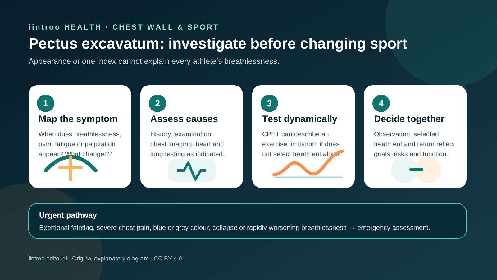
Pectus excavatum can affect exercise in different ways. Symptoms, examination and selective CPET—not appearance or one index—should guide sport decisions.
Key points
- Chest appearance and a Haller index alone do not explain every episode of breathlessness or determine sport eligibility.
- Current multidisciplinary guidance recommends cardiopulmonary exercise testing for severe pectus excavatum with exercise intolerance, alongside evaluation for other causes.
- Surgery can improve symptoms and some measured outcomes in selected patients, but evidence is non-randomized, heterogeneous and not a performance guarantee.
Start with the athlete's symptoms, not the chest photograph
This guide is for athletes with pectus excavatum, families, coaches and clinical teams. Pectus excavatum is a depression of the front chest wall with wide variation in depth, symmetry, age of presentation and effect. Some athletes have no functional limitation; others report exertional breathlessness, chest discomfort, early fatigue, palpitations or reduced confidence. Appearance does not reveal whether the heart, lungs, conditioning, asthma, anaemia, anxiety, another condition or several factors are contributing.
Ask what changed, at which intensity, in which position and with which associated symptoms. Compare normal training with racing, heavy lifting, prone or compressed positions and growth-related changes. Fainting during exercise, severe chest pain, blue or grey colour, rapidly worsening breathlessness or collapse needs urgent assessment. Persistent exertional symptoms deserve qualified evaluation rather than a coach diagnosing deconditioning or telling the athlete to train through them.
Use imaging, heart and lung assessment—and CPET for the right question
The 2024 joint specialist-society guideline recommends cross-sectional chest imaging when evaluating suspected exercise intolerance, plus echocardiography and lung-function testing to look for anatomy and alternative explanations. It recommends cardiopulmonary exercise testing, or CPET, in severe pectus excavatum with exercise intolerance because a dynamic test can describe oxygen uptake, heart-rate response, ventilation and exercise limitation together. These investigations are interpreted as a set; a normal resting test does not automatically settle an exertional complaint.
The Haller index describes chest shape on imaging but is not a self-contained measure of symptoms, cardiac compression or eligibility. Technique, breathing phase, correction index, age and the relationship between the sternum and heart matter. CPET also requires adequate effort and appropriate reference values. A result below a predicted threshold can support further assessment, but it does not identify one cause by itself or turn an athlete into a surgical candidate without the full clinical context.
Discuss treatment benefits without promising a faster athlete
Treatment can include observation, posture and conditioning support, vacuum-bell therapy in selected people, psychological support and surgical repair. The choice depends on age, anatomy, symptoms, objective findings, goals, risks and local expertise. A 2022 single-centre adult cohort reported improved CPET measures several years after minimally invasive repair, and a UK review found many studies reporting physiological or quality-of-life benefit. Both bodies of evidence are dominated by selected, non-randomized patients and heterogeneous tests.
A 2025 meta-analysis of 15 non-randomized studies found serious or critical risk of bias in every included study and did not demonstrate a clinically significant improvement in peak oxygen uptake. A larger 2026 retrospective cohort reported improved paired CPET outcomes, but it still cannot establish what would have happened without surgery and disclosed relevant author conflicts. Surgery may be appropriate for selected clinical reasons; it should not be sold as guaranteed performance enhancement or as the only valid response to body-image distress.
Keep training specific, progressive and connected to follow-up
While assessment is under way, the clinician may recommend modifying intensity or the activity that provokes symptoms rather than stopping all movement. Record session type, duration, symptoms, recovery and any heart-rate or power context without treating a wearable as a diagnostic device. After any procedure, return must respect wound healing, bar position, pain, breathing, shoulder and trunk function, contact risk and the surgeon's restrictions. Swimming, rowing, lifting and collision sport place different demands on the chest wall.
Reassessment is appropriate when symptoms change, growth is rapid, a new sport increases contact or pressure, or performance falls without an obvious training explanation. Coaches should support privacy and avoid comments that turn chest shape into a measure of effort or masculinity. There is no universal Haller index, CPET value, exercise heart rate or post-operative week that clears every athlete. This page is general education, not diagnosis, imaging interpretation, treatment selection, exercise prescription or personal return-to-sport clearance.