
Peroneal tendon problems in athletes: when lateral ankle pain is not another sprain
Persistent posterolateral ankle pain or snapping can reflect peroneal tendon disease. Diagnosis and return depend on the specific tissue problem and sport demand.
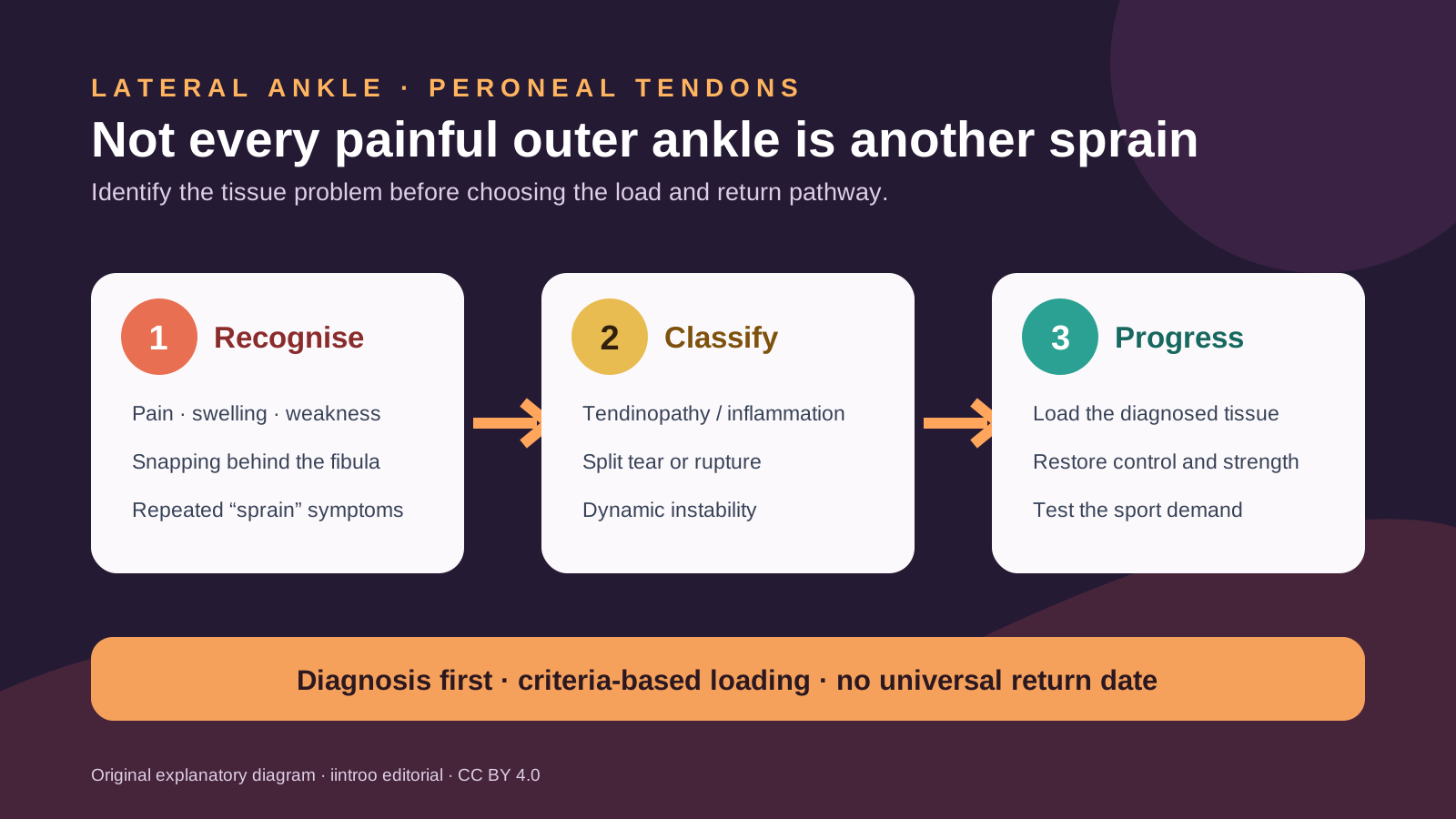
Key points
- Peroneal tendinopathy, tears and tendon instability can resemble a recurrent lateral ankle sprain but require different assessment and management.
- MRI and ultrasound can both help; dynamic ultrasound is particularly useful when snapping or subluxation occurs during movement.
- Running and return to sport should be criteria-based; published rehabilitation and surgical studies are heterogeneous and do not provide one reliable date.
Look beyond a repeated ankle-sprain label
This guide is for runners, skiers, footballers, court-sport athletes and others with pain or snapping behind the outer ankle bone. The peroneus brevis and longus tendons help evert and stabilize the foot. They can develop tendinopathy or tenosynovitis, split or full-thickness tears, or instability in which a tendon moves abnormally around or within the retromalleolar groove. These conditions may follow an ankle sprain, sudden forceful dorsiflexion and eversion, repetitive uneven-ground loading or another injury, but symptoms and mechanisms overlap.
Pain, swelling, weakness or tenderness along the posterolateral ankle deserves assessment when it persists, repeatedly returns or fails to behave like an uncomplicated sprain. A visible or felt snap with movement, a sense that something moves behind the fibula, recurrent giving way or loss of push-off strength increases concern for instability or a tear. Inability to bear weight, marked deformity, rapidly increasing swelling, neurovascular change or acute severe pain after trauma needs timely clinical evaluation and appropriate fracture screening.
Match imaging to the suspected problem
The ESSKA-AFAS consensus states that both MRI and ultrasound are appropriate after clinical examination. MRI can show tendon morphology, associated ligament or cartilage injury and anatomy around the groove. Ultrasound can examine the tendons in real time while the foot moves, which is valuable for subluxation or intrasheath instability that may not appear on a static scan. A 2025 diagnostic meta-analysis found promising pooled accuracy for ultrasound, but included studies were small and heterogeneous, so no percentage guarantees a correct result in every setting.
Imaging must be interpreted with symptoms and examination. Position-dependent movement can sometimes be normal, while a report can miss a dynamic lesion or identify a change that is not the source of pain. A 2025 tendoscopy systematic review included only eight studies and 190 patients, largely surgical series. It supports the idea that clinically important pathology may remain difficult to define, but it does not justify invasive investigation for every sore ankle. Specialist review is appropriate when suspicion remains high despite initial care or imaging.
Rehabilitation depends on tendinopathy, tear or instability
For many tendinopathy presentations, initial care may include temporary load modification, restoration of ankle motion, progressive calf and peroneal strength, balance and control, and attention to associated ankle instability or foot mechanics. The exact plan depends on irritability, sport, terrain, footwear and whether another injury is present. Repeated aggressive stretching, massage over a snapping tendon or testing uneven-ground running through escalating pain can worsen symptoms and cannot determine whether a retinaculum is injured.
Acute dislocation, recurrent symptomatic instability, substantial tears and high-demand elite sport may lead to a surgical discussion, but procedure choice belongs with a foot-and-ankle specialist. Reviews report favourable outcomes in many surgical cohorts, yet much of the literature is level IV evidence, rehabilitation protocols vary and not every athlete returns to the same level. Treatment should address the diagnosed structure rather than using a generic tendon protocol or copying another athlete's immobilization schedule.
Return by function and sport exposure, not a countdown
Progression generally moves from comfortable daily walking and controlled strength toward single-leg calf work, balance, hopping, acceleration, cutting, uneven terrain and sport-specific fatigue as permitted. The international consensus recommends that running after surgery should depend on the athlete's pre-injury condition, single-heel-rise capacity, overall strength, neuromuscular control and proprioception rather than time alone. The relevant test battery differs for a trail runner, skier, football winger or dancer.
Pain, swelling, snapping, weakness or instability during a stage—or a clear next-day increase—should pause progression and prompt review. Return after a repaired superior peroneal retinaculum follows different loading rules from nonoperative tendinopathy, so online timelines are not interchangeable. This page cannot diagnose a tear, choose MRI or ultrasound, prescribe immobilization, recommend surgery or provide clearance. The evidence supports precise diagnosis and criteria-based loading; it does not support a universal brace, injection or return date.