
Popliteal artery entrapment in runners: investigate repeatable calf symptoms
Repeatable calf pain, weakness or numbness at hard effort can be vascular. Dynamic testing helps distinguish popliteal entrapment from other leg problems.
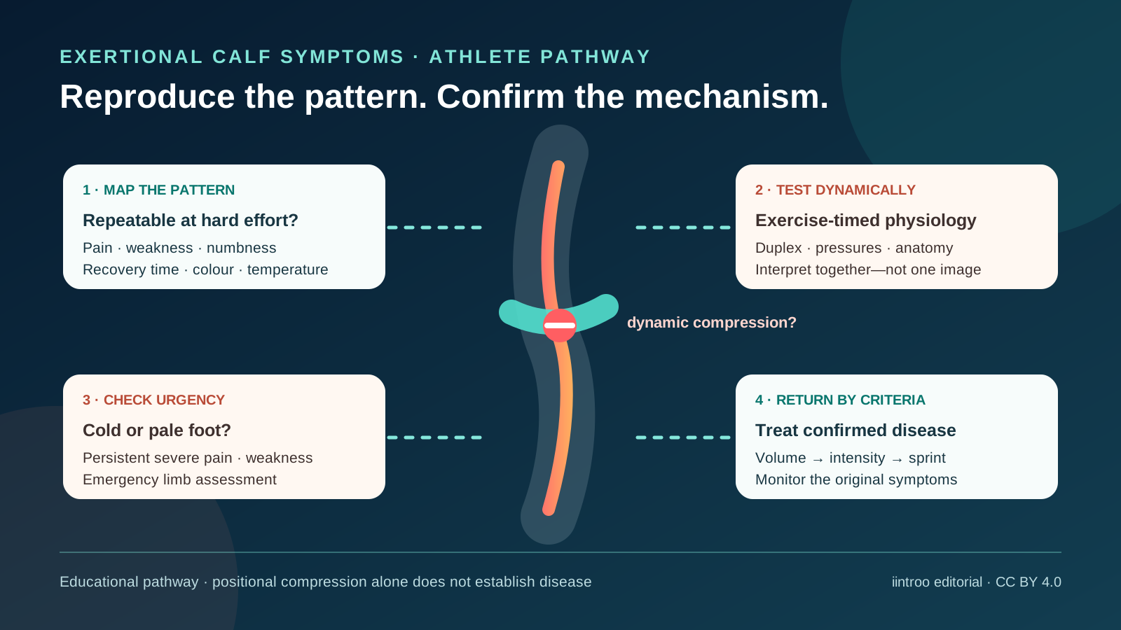
Key points
- Popliteal artery entrapment syndrome can cause reproducible calf pain, cramping, weakness or altered sensation during exercise in otherwise healthy athletes.
- Resting pulses and imaging may be normal; history, examination and vascular tests timed to symptom-provoking activity are interpreted together.
- Positional artery compression also occurs in people without symptoms, so one provocative ultrasound finding cannot establish the diagnosis or justify surgery.
Recognize a vascular pattern without labelling every calf pain
This guide is for runners, footballers and other athletes with calf pain, tightness, weakness, heaviness or altered sensation that appears predictably at a certain intensity and eases after stopping. Popliteal artery entrapment syndrome occurs when structures behind the knee compress the artery. Anatomical forms involve an unusual relationship between artery and muscle or tendon; functional entrapment can occur without that fixed anomaly, often in highly trained young adults. Mileage or large calf muscles alone do not diagnose it.
The differential diagnosis is broad. Chronic exertional compartment syndrome, bone stress injury, nerve entrapment, tendon or muscle injury, venous disease and referred pain can produce overlapping symptoms, and more than one problem may coexist. Record which leg and region are affected, the speed or duration that triggers symptoms, whether power falls, how quickly recovery occurs and whether foot colour, temperature or sensation changes. Note terrain, footwear and the exact point at which the symptoms stop performance. A normal easy run does not exclude a problem that only appears near maximal effort, and a training diary can help a clinician reproduce the relevant workload safely.
Match the test to the exercise-triggered problem
Evaluation combines history and vascular examination with tests selected by a sports and vascular team. The American College of Radiology considers duplex Doppler, contrast-enhanced magnetic-resonance angiography and CT angiography appropriate initial imaging options for suspected entrapment. Ankle-brachial pressures before and immediately after symptom-provoking exercise may add information. Timing matters because blood flow can normalize rapidly once activity stops, leaving a resting study falsely reassuring.
Dynamic ultrasound can examine the artery during loaded ankle movements or standing, but compression is not automatically disease. A 2024 controlled study found distal popliteal occlusion during prone plantar flexion in many healthy controls and recreational athletes, illustrating the false-positive problem. Symptoms, location of compression, haemodynamic change and cross-sectional anatomy must agree. A single bedside manoeuvre, consumer circulation device or screenshot of an ultrasound should not determine treatment.
Escalate acute limb signs and avoid self-testing through danger
A suddenly cold, pale or blue foot, severe pain, new weakness or numbness, absent pulse or symptoms that do not settle after stopping can indicate acute limb ischaemia and needs emergency assessment. Do not massage the leg, repeat sprints to prove the pattern or wait for a routine sports clinic. Progressive arterial wall damage, thrombosis or embolisation is uncommon but is why persistent exertional vascular symptoms deserve timely evaluation rather than months of load modification alone.
For stable symptoms, reducing the provoking workload while assessment proceeds is different from prescribing permanent rest. Training alternatives should not recreate calf compression or conceal worsening signs. Medication, injections or surgery should not be selected from an online description. Recent reviews emphasize that diagnostic standards and comparative treatment evidence remain limited; misdiagnosis can lead either to delayed care or an unnecessary procedure.
Return from the confirmed diagnosis, not a generic timeline
Management depends on anatomical versus functional disease, artery damage, symptom burden, sport goals and competing diagnoses. Surgical decompression is used for selected symptomatic cases. In a single-centre retrospective series of 73 athletes and 125 limbs, 82.4% returned to their previous competitive level after a standardized surgical approach; recurrence still occurred and the cohort cannot prove that the same operation is best for all athletes. Smaller series and differing definitions limit certainty.
Return should restore walking and daily function, then running volume, intensity, hills, sprinting and sport-specific change of direction while monitoring for the original reproducible symptoms. Vascular findings, wound or muscle recovery and any coexisting compartment problem affect progression. Coaches should know which symptoms require an immediate stop and which follow-up is planned. This page cannot diagnose entrapment, interpret imaging, recommend botulinum toxin or surgery, or provide a universal return date. The safe sequence is symptom reproduction under qualified assessment, multimodal confirmation, individualized treatment and criteria-led reloading.