
Midfoot pain in athletes: a navicular stress injury is easy to miss and risky to ignore
Focal midfoot pain can signal a high-risk navicular bone stress injury. Early assessment, appropriate imaging and staged return help protect healing.
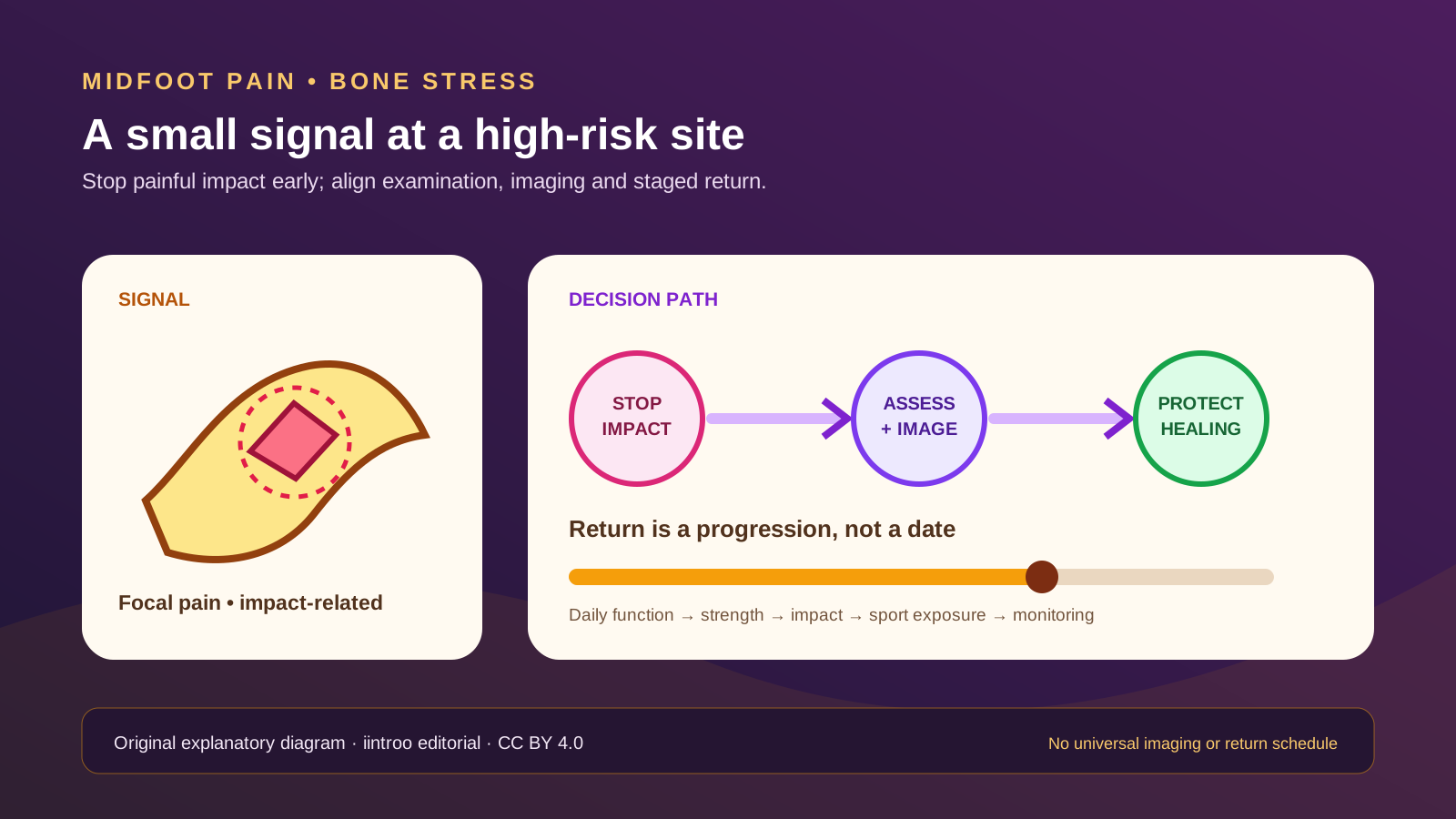
Key points
- The tarsal navicular is considered a high-risk bone stress site because delayed healing and non-union can occur.
- Plain radiographs may not show an early stress injury; imaging choice and interpretation belong with a clinician who understands the examination and timing.
- Return should follow demonstrated healing, restored function and gradual sport-specific loading, not a universal calendar or a pain-masking test.
Do not run through a small but persistent midfoot signal
This guide is for runners, jumpers, court athletes, dancers, military athletes and their support teams. Navicular bone stress injury often develops without one dramatic event. Pain may be vague at first, worsen with running or hopping and settle at rest, while swelling can be absent. Because the navicular sits deep in the midfoot, athletes may call it an ankle ache, lace pressure or a general foot niggle. Repeatedly testing it with hard sessions can increase load before the injury is named.
Stop painful impact and seek timely sports-medicine assessment when focal midfoot pain persists, recurs with loading or changes gait. Inability to bear weight, marked swelling, deformity, neurovascular symptoms, fever, a wound or acute high-energy trauma needs urgent evaluation. The navicular is a high-risk site in international bone-stress consensus work and return-to-sport reviews because complication rates are higher than at many lower-risk locations. That classification is a reason for early assessment, not a prediction that every athlete needs surgery.
Combine examination, training history and the right image
A clinician should ask about the exact pain location, growth and menstrual history when relevant, recent changes in running or jumping, surfaces, footwear, previous bone injuries, nutrition, sleep, medications and other medical conditions. Focal tenderness over the dorsal navicular can raise suspicion, but no single touch or hop test confirms the diagnosis. Differential diagnoses include joint, tendon, ligament and other bone injuries, so an athlete should not self-diagnose from a body map.
Early radiographs can be normal. The 2025 foot-and-ankle sports consensus discusses MRI findings such as bone oedema and CT characterization of a fracture, with the imaging sequence shaped by the clinical question. More scanning is not automatically better, and incidental findings must be interpreted in context. The important practical point is that a negative early X-ray does not safely rule out navicular stress injury when the history and examination remain concerning.
Protect healing and investigate contributors
Management varies with the injury grade, fracture characteristics, healing, competition level and athlete preference. It may include protected or non-weight-bearing activity, rehabilitation or surgery after shared decision-making. The consensus provides broad pathways for elite athletes, but those recommendations should not be converted into a universal boot duration or operation rule. Continuing impact because cycling or daily walking feels comfortable can still conflict with the treating plan.
At the same time, review contributors rather than blaming one session. Rapid load increases, inadequate recovery, low energy availability, menstrual disturbance, low bone density, previous bone stress injury and nutritional or medical factors may matter in different combinations. The 2025 Female Athlete Triad update and broader bone-stress consensus support structured assessment, but sex alone does not diagnose a cause. Supplements should not be started from a social-media checklist; indicated blood tests and nutrition care require qualified interpretation.
Return through function, impact and sport exposure
Return is a continuum: maintain safe conditioning within restrictions, restore comfortable daily weight-bearing, rebuild calf and foot capacity, then add impact and sport-specific tasks when the treating team agrees healing is adequate. Monitor symptoms during activity and over the next day. Running distance, speed, hills, jumping, cutting and competition should not all be reintroduced together. A single pain-free hop cannot reproduce accumulated load or prove bony union.
Published return times vary with injury severity, management and population. The 2025 elite-athlete consensus reports typical outcomes after surgery, while later commentary stresses limits in the evidence base; neither creates a deadline for a recreational or adolescent athlete. The safer message is to identify the site early, protect the biology of healing and progress only when clinical, imaging and functional information align. This page cannot select imaging, prescribe weight-bearing, recommend surgery or clear a return.