
Gluteal tendinopathy in running: rebuild lateral-hip load before mileage
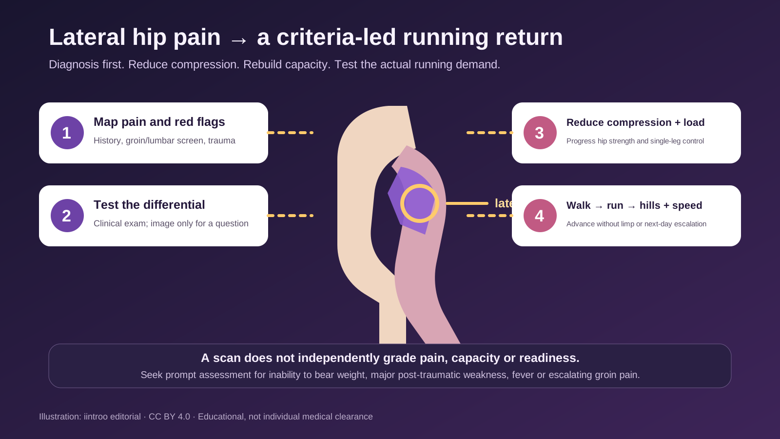
Lateral hip pain may reflect gluteal tendinopathy. Diagnosis, reduced compression and progressive hip loading should come before a full running return.
Key points
- Gluteal tendinopathy is one cause of pain around the greater trochanter; hip-joint, lumbar, fracture and other tendon problems can produce a similar location.
- Clinical examination drives diagnosis, while imaging may clarify a tear or alternative cause but does not grade pain or disability by itself.
- Education and progressive exercise have the strongest conservative evidence; return rebuilds walking, single-leg control, running volume and sport intensity in sequence.
Map lateral-hip pain before calling it bursitis
This guide is for runners, cyclists, field and court athletes with pain over the outside of the hip, often worse with running, stairs, single-leg stance, side-lying or crossing the legs. Greater trochanteric pain syndrome is an umbrella term; gluteus medius or minimus tendinopathy is a common driver, while bursal irritation may coexist. The painful location alone does not identify the tissue. Hip osteoarthritis, femoral stress injury, lumbar referral, nerve problems, acute tendon tear and other regional conditions require different decisions.
A clinician asks about onset, recent changes in running volume, hills, speed, surface, sleep positions and symptoms below the knee or in the groin. Examination can combine local palpation, resisted hip abduction, single-leg loading, gait and hip or lumbar screening. Sudden inability to bear weight, major weakness after trauma, fever, night pain with systemic illness or escalating groin pain needs prompt assessment. The athlete should not repeatedly run through a limp to prove that the pain warms up.
Use imaging to answer a question, not to score the athlete
A 2024 diagnostic systematic review found that a sequence of local palpation and resisted-abduction testing can meaningfully change the probability of greater trochanteric pain syndrome, although no single test is perfect. Ultrasound or MRI may help when the diagnosis is uncertain, symptoms persist, significant weakness suggests a tear or an intervention is being considered. Radiographs can assess bone and joint alternatives. Imaging should follow a clinical question rather than act as a routine badge of severity.
A 2025 analysis of 204 trial participants found tears and other tendon changes were common on MRI, but imaging severity was not associated with pain, function or disability. That result does not mean imaging is useless or every tear is harmless; it shows why a scan cannot independently prescribe rest, surgery or return. Runner-specific evidence remains smaller than the general middle-aged clinical literature, and women are heavily represented in many studies. Training decisions therefore need direct functional testing and the athlete's response to load.
Reduce compression while rebuilding tendon capacity
Contemporary management uses education to reduce avoidable tendon compression and organise load, then progressive exercise to restore capacity. Early changes may include avoiding prolonged hip adduction, changing painful side-lying positions and temporarily reducing hills, speed or long runs rather than stopping all movement. Strength work usually progresses from tolerable hip-abductor loading to heavier and more functional single-leg tasks. The exact exercises, range and dose must be individualized; pain monitoring is a decision aid, not a universal traffic-light rule.
Systematic reviews published in 2024 and 2025 support education plus exercise as the core conservative approach, with better long-term global improvement than corticosteroid injection in the available trials. Injection and shockwave evidence varies by outcome and follow-up, and platelet-rich plasma findings are not strong enough to promise a superior result. Apparent short-term relief does not demonstrate restored tendon capacity. Sleep, general health, bone risk and other hip diagnoses may also influence progress, especially when symptoms are prolonged or the athlete is older.
Return from walking capacity to running density and speed
A return plan first restores comfortable daily walking, stairs and controlled single-leg loading without a meaningful next-day flare. It can then add short flat run-walk exposures, continuous easy running, longer duration, hills, speed and sport-specific cutting or jumping. Track lateral-hip pain, gait quality, strength, confidence and recovery across several sessions. Cadence, footwear or surface changes may help an individual problem, but none is a universal correction and several variables should not be changed at once.
Return is not defined by a normal scan, a fixed number of weeks or one pain-free session. Recurrent night pain, increasing weakness, a limp or inability to progress despite a well-delivered programme warrants reassessment. This article cannot diagnose the source of hip pain, prescribe exercises, recommend an injection or clear competition. It provides a framework for accurate diagnosis, compression-aware load management and gradual exposure to the mileage, speed, terrain and direction changes the athlete actually needs.